TF-Scan – non-invasive
tear film assessment

Diagram 1:
Schirmer’s test (Source, W. Sickenberger)

Diagram 2:
BUT-Test
Dry eye
Dry Eye Syndrome is the result of inadequate wetting of
the ocular surface. Dry eyes have numerous causes. Tear
film production and composition play a prominent and
important role. A disorder of the lipid layer leads to
faster evaporation of the tear film and thus to dry eyes
with the aforementioned complaints. Symptoms include
stinging and scratching eyes, foreign body eye sensation,
redness, increased sensitivity to light as well as contact
lens and cosmetic intolerance. Working long hours at
a monitor aggravates these problems in particular.
Tear film assessment
In terms of tear film assessment one can generally distinguish
between invasive and non-invasive and between quantitative
and qualitative methods. Methods are designated as invasive
when they involve eye contact or application of fluorescein,
for example. In contrast, noninvasive methods are contact-free.
Quantitative tear film analysis assesses the amount of
tear film whereas qualitative tear film assessment provides
information on tear film stability as well as tear film
composition. The goal is to be able to determine the tear
film quantity and quality non-invasively. Non-invasive
measurements of the tear film do not irritate the eye,
thus delivering more reliable results. Moreover, non-invasive
methods are considerably more patient-friendly.
Schirmer's test (Diagram 1) is often used in practice
to determine tear film quantity. This test involves hanging
a strip of filter paper inside the lower lateral conjunctival
sack. The litmus test strip becomes saturated with lacrimal
fluid. The assessment is made after 5 minutes.
The invasive BUT test (Diagram 2), which entails applying
fluorescein, is quite widely used for assessing tear film
quality. The BUT test can be used to determine the break-up
time of the tear film up to the point where dry patches
occur. The longer the tear film needs to break up, the
more stable it is. Both tests are invasive, however, and
not always pleasant for the patient.
Qualitative assessment by using the OCULUS Keratograph
In close collaboration with JENVIS Research at the Jena
University of Applied Sciences the TF-Scan (tear film
scan) was developed for the OCULUS Keratograph (topographer).
With this new software the tear film can be assessed
qualitatively and quantitatively. Both measurements are taken non-invasively and thus in a more sanitary and patient-friendly way.

Diagram 3: Edge shift of placido rings (Source: D.Wiedemann, JENVIS Research Institute

Diagram 4: Automatic detection and evaluation |

Diagram 5: Color visualization of break-up time |
The break-up time of the tear film (quality) can be determined
using the time-tested NIBUT procedure (non-invasive break-up
time). Changes in the projected placido rings (edge shift
of placido rings) indicates the break-up time of the tear
film (Diagram 3 and 4). This evaluation is made automatically
and the break-up time is represented in color. Dry patches
which break up early on are marked in red. Color representation
facilitates patient consultation considerably (Diagram
5).
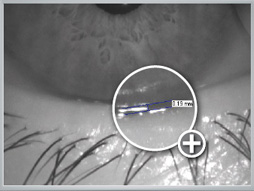
Diagram 6: Measured tear meniscus height
Quantitative assessment by using the OCULUS Keratograph
In addition the quantity of the tear film can be assessed
non-invasively. To do this the tear meniscus height is
determined. In comparison to Schirmer's test this method
is much more pleasant for the patient. A tear meniscus
height of 0.2 mm or less indicates that the amount of tear film
is too small.
The tear meniscus height (quantity) can be measured and
assessed quickly and easily after the data has been recorded
using the keratograph software (Diagram 6).